Mi1 [FBbt_00003776]

Mi1
ID: FBbt_00003776
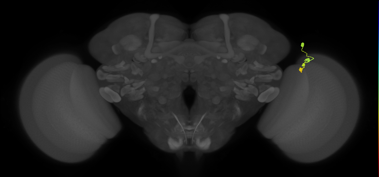
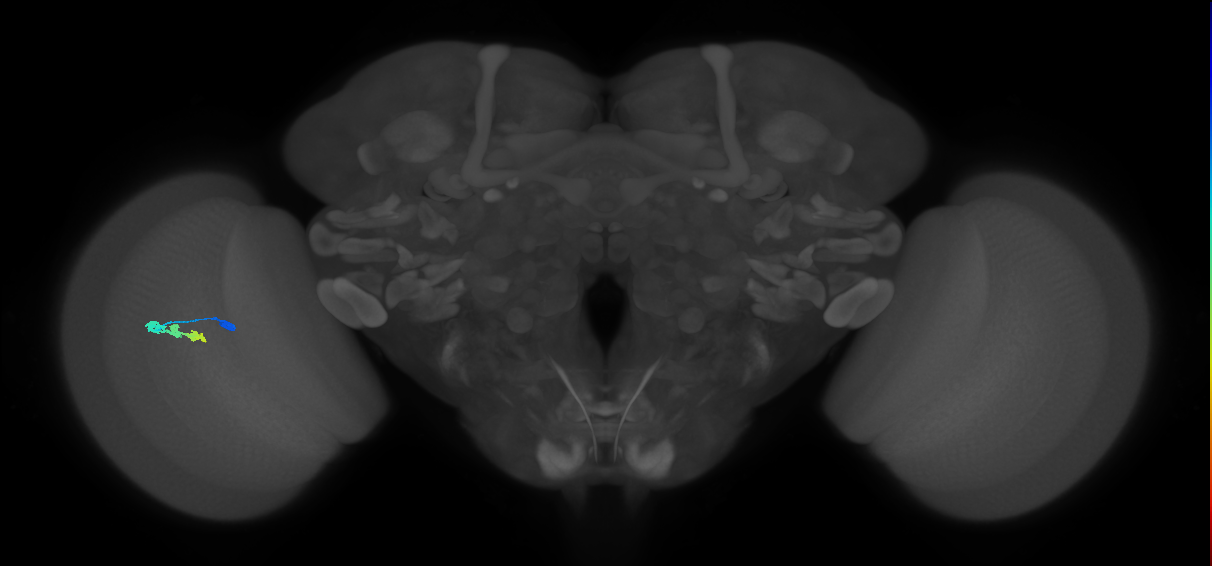
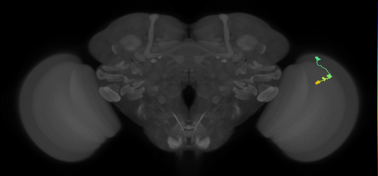
Unicolumnar medullary intrinsic neuron with bushy, fine arborizations in medulla layers M1, M5 and M9-10 (Fischbach and Dittrich, 1989; Kind et al., 2021). The projection of this neuron branches at the inner-face of the medulla to form two to three varicose recurrent terminal specializations that extend back up to the inner border of layer M8 (Fischbach and Dittrich, 1989; Morante and Desplan, 2008). In central (non-dorsal margin) columns it receives input from an R8 photoreceptor cell, but connections are substantially weaker in dorsal margin columns (Kind et al., 2021). Pre-synaptic terminals are present mainly in medulla layers M9-M10, but also in all other layers where neurites extend laterally (Pankova and Borst, 2017). It receives input from lamina monopolar neurons L1 and L5 and centrifugal neuron C2 (Takemura et al., 2013). It outputs to T4 neurons and transmedullary neuron Tm3a (Takemura et al., 2013). It is a cholinergic neuron (Hasegawa et al., 2011; Pankova and Borst, 2017). There is usually one of these cells per optic column (Nern et al., 2025).
Pre-synaptic terminals were assessed by labelling with a UAS-Synaptotagmin reporter (P{UAS-HA-syt} - FBtp0015803). The neurotransmitter was assessed by labelling cells with an anti-ChAT antibody (Hasegawa et al., 2011). The morphology of the terminals was judged from figures in Fischbach and Dittrich (1989) as assessed by FlyBrain Neuron DB. Connectivity and morphology in the medulla were assessed by electron microscopy reconstruction from 7 columns (Takemura et al., 2013).
Open in VFB 3D Browser →- Notch ON hemilineage secondary neuron
- adult cholinergic neuron
- medulla intrinsic columnar neuron
- optic lobe narrow field columnar neuron
- visual system neuron
Relationships
- develops from: immature medulla intrinsic neuron Mi1
- receives synaptic input from neuron: centrifugal neuron C2, lamina monopolar neuron L1, lamina monopolar neuron L5
- receives synaptic input in region: medulla layer M1, medulla layer M5, medulla layer M6
- sends synaptic output to cell: T4 neuron, transmedullary neuron Tm3
- sends synaptic output to region: medulla layer M1, medulla layer M10, medulla layer M5, medulla layer M6, medulla layer M9
Alternative Names
| Synonym | Scope | Reference |
|---|---|---|
| small field unilateral tristratified neuron | related synonym | |
| medullary intrinsic neuron 1 | exact synonym | |
| medullary intrinsic neuron Mi1 | exact synonym | |
| Mi1 | exact synonym | Nern et al., 2025 |
| Mi1 | exact synonym | Özel et al., 2021 |
| Sut | related synonym | |
| Mi1 | exact synonym | Schlegel et al., 2024 |
Feedback
Was this page helpful?
Glad to hear it! Please tell us how we can improve.
Sorry to hear that. Please tell us how we can improve.