T2a [FBbt_00003729]


T2a
ID: FBbt_00003729
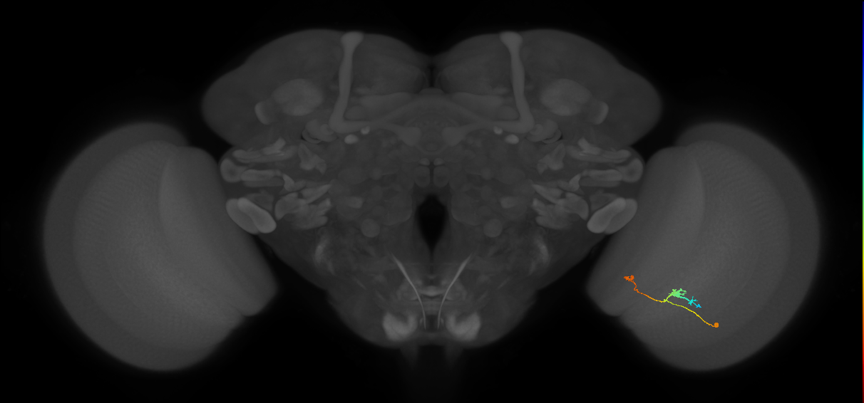
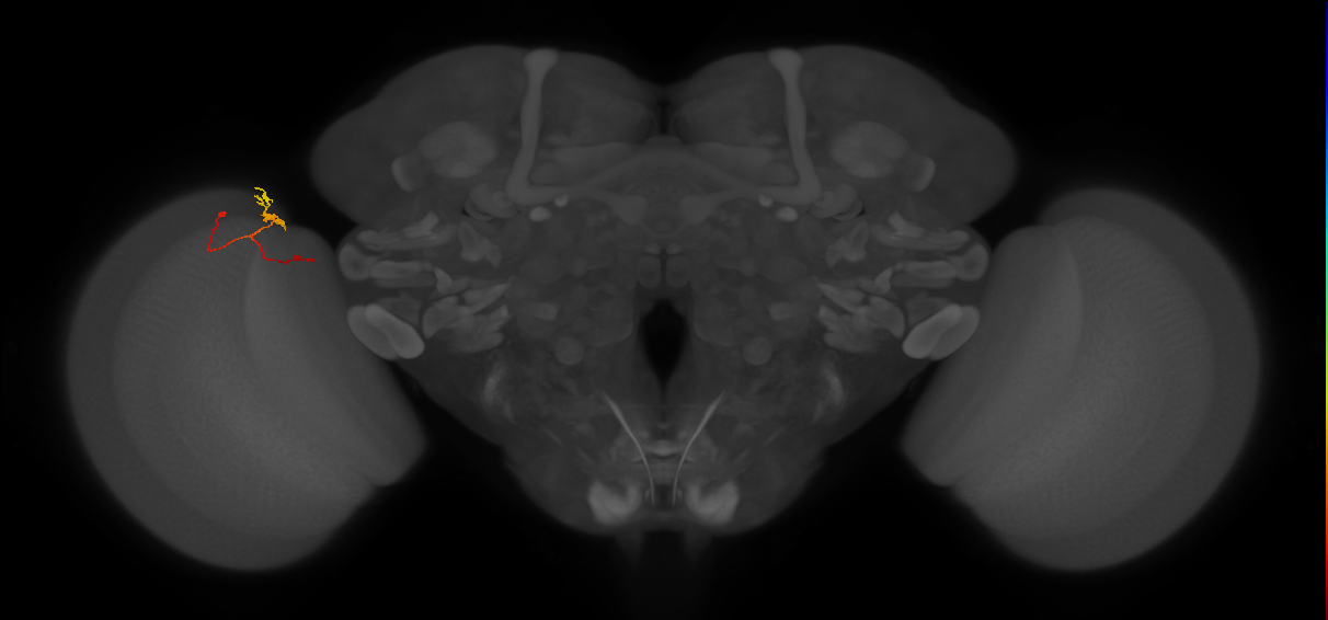
T neuron with its soma posteriorly adjacent to the gap between the medulla and lobula plate and a cell body fiber that projects along the proximal surface of the medulla before branching in the second optic chiasm (Fischbach and Dittrich, 1989; Shinomiya et al., 2019). One branch projects into a medulla column, where it forms a bushy, fine, arborization in medulla layer M9 and then projects through the rest of the medulla column, bifurcating in medulla layer M5 and forming fine arbors throughout layers M1-4. The other branch forms a terminal arborization in lobula layer 3 that is much wider than a single column and has bleb-type terminal branches.
Pre- versus postsynaptic innervation judged by scoring of terminal morphology from figures in Fischbach and Dittrich (1989) as assessed by FlyBrain Neuron DB.
Open in VFB 3D Browser →Relationships
- has soma location: cell body rind of adult posterior lobula plate
- receives synaptic input in region: medulla layer M1, medulla layer M10, medulla layer M2, medulla layer M3, medulla layer M4, medulla layer M5, medulla layer M8, medulla layer M9
- sends synaptic output to region: lobula layer 3
Alternative Names
| Synonym | Scope | Reference |
|---|---|---|
| T neuron T2a | exact synonym | |
| T2a | exact synonym | Nern et al., 2025 |
| T2a | exact synonym | Özel et al., 2021 |
| T2a | exact synonym | Schlegel et al., 2024 |
Feedback
Was this page helpful?
Glad to hear it! Please tell us how we can improve.
Sorry to hear that. Please tell us how we can improve.