ON and OFF motion detection circuit
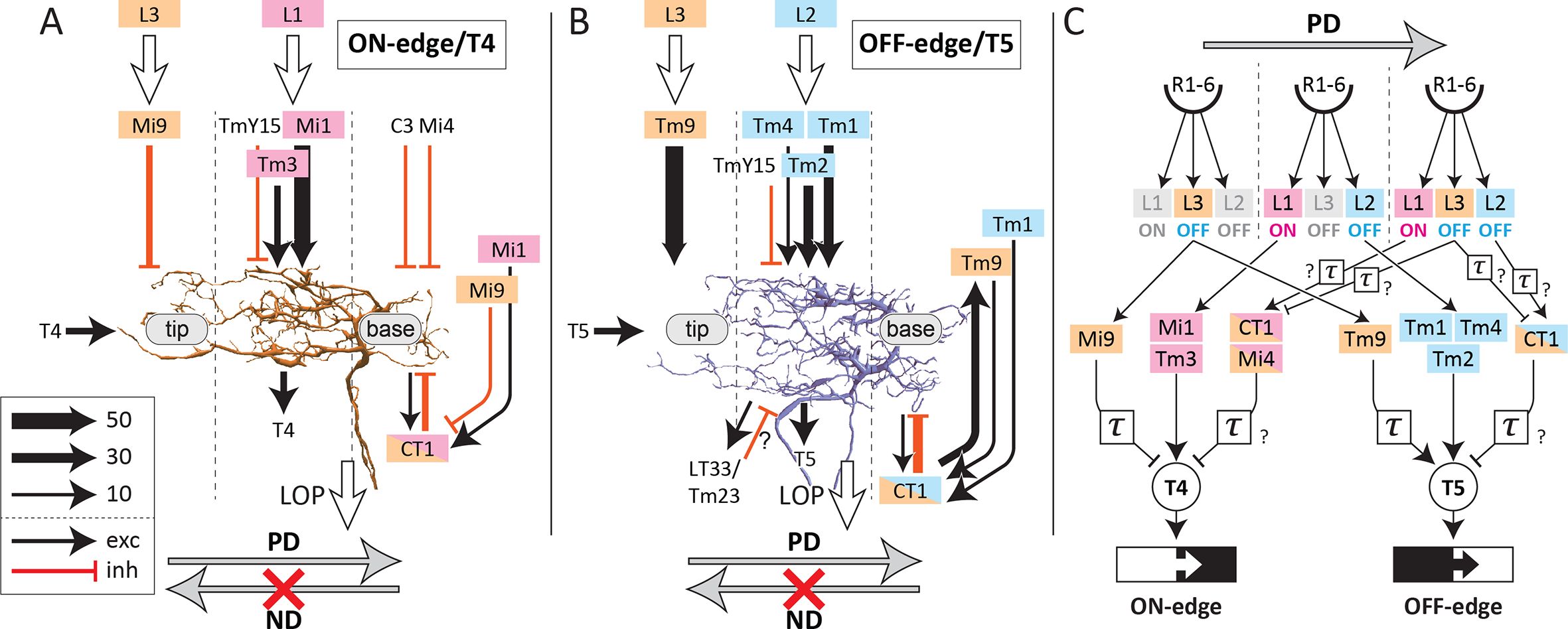
Schematic wiring of the ON-edge (T4) and OFF-edge (T5) motion-detection circuits, from Figure 6 of Shinomiya et al. (2019). Panels A and B show the columnar inputs that converge on T4 and T5 dendrites — Mi1, Mi4, Mi9, Tm3, TmY15, C3 (and CT1 feedback) for T4; Tm1, Tm2, Tm4, Tm9, TmY15 (and CT1) for T5 — with arrow weights indicating relative synapse counts. Panel C is the canonical summary cartoon: photoreceptors R1-6 → lamina monopolar cells L1/L2/L3 → medulla cells (Mi1/Mi4/Mi9/Tm3 driving T4 ON-edge; Tm1/Tm2/Tm4/Tm9 driving T5 OFF-edge) → T4/T5 → lobula plate. Click a labelled neuron type or neuropil to open the corresponding term in Virtual Fly Brain.
Categories:
https://doi.org/10.7554/eLife.40025
Figure reproduced under CC-BY 4.0 from Shinomiya K, Huang G, Lu Z, Parag T, Xu CS, Aniceto R, Ansari N, Cheatham N, Lauchie S, Neace E, Ogundeyi O, Ordish C, Peel D, Shinomiya A, Smith C, Takemura S, Talebi I, Rivlin PK, Nern A, Scheffer LK, Plaza SM, Meinertzhagen IA (2019). Comparisons between the ON- and OFF-edge motion pathways in the Drosophila brain. eLife 8:e40025.
Feedback
Was this page helpful?
Glad to hear it! Please tell us how we can improve.
Sorry to hear that. Please tell us how we can improve.
Last modified May 8, 2026: Anatomy diagrams: rescale image-map coords to displayed pixel space (3451b92)